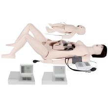
The female pelvic model, a type of Human Anatomical Model, is a detailed and accurate representation of the muscles and organs that make up the female pelvis. It is designed to help medical students and professionals, particularly those involved in Obstetrics And Gynecology Skills training, understand the structure and function of these important parts of the female reproductive system.
The model typically includes the pelvic bones, uterus, ovaries, fallopian tubes, bladder, rectum, and various muscles and ligaments. These structures are carefully crafted to match the size, shape, and texture of their real-life counterparts, allowing users to study them in detail and gain a deeper understanding of their anatomy.
The model may also feature removable parts, such as the uterus or bladder, to allow for closer examination and study. This feature makes it an invaluable tool in Clinical Skill Training Models, as it provides a hands-on approach to learning.
Some models may also include additional features, such as color-coding or labeling of different structures, to aid in learning and identification. This is particularly useful in Nursing Skill Training Models, where understanding the exact location and function of each structure is crucial.

Overall, the female pelvic model is an essential tool for anyone studying or working in the fields of gynecolo
gy, obstetrics, or pelvic health. Whether used as a standalone Human Anatomical Model or as part of a comprehensive Clinical Skill Training Model, it helps users develop a deeper understanding of this complex and important part of the human body.
Features:
Model is designed to demonstrate female pelvic muscles and organs.