A urinary system anatomy model can be a valuable tool for visualizing and studying the structures involved. These models typically include the kidneys, ureters, bladder, and urethra, allowing for a comprehensive exploration of the urinary system's components.
With a urinary system anatomy model, you can examine the intricate details of each organ and understand their functions. The kidneys, bean-shaped organs located on either side of the spine, filter waste products and excess fluid from the blood. The ureters, thin tubes connected to the kidneys, transport urine to the bladder. The bladder, a muscular sac located in the pelvis, stores urine until it is expelled through the urethra.
Using a urinary system anatomy model, you can explore the relationships between these organs and study how they work together to maintain homeostasis. You can also use the model to learn about common urinary system conditions and treatments, such as kidney stones, urinary tract infections, and urinary catheterization techniques.
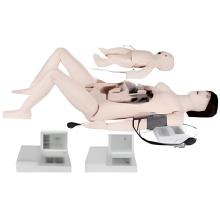
For medical professionals and students studying urology or related fields, it's essential to have hands-on training using specialized models. For example, first aid skill training models, such as the Full Body Trauma Manikin or the Mattress Sutures Model, provide practical experience in emergency situations. These models simulate real-life scenarios and enable learners to develop essential skills in Trauma Care.
Additionally, there are specific models designed for advanced medical procedures. The Intravenous Injection Arm and the Intramuscular Injection Model are valuable tools for learning proper injection techniques. These models allow practitioners to practice these skills in a controlled environment before performing them on real patients.
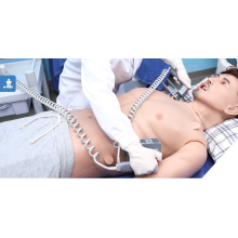
Simulation models also play a crucial role in training healthcare professionals in life-saving techniques. The Electronic Cpr Manikin, for example, provides realistic simulations of cardiac arrest scenarios, allowing learners to practice CPR and ACLS (Advanced Cardiac Life Support) training effectively. Similarly, the Aed Training models help individuals develop the necessary skills to operate automated external defibrillators in emergency situations.

In summary, the use of anatomical models and training manikins greatly enhances medical education and skill development. These models offer a hands-on approach to learning and provide a safe environment for medical professionals and students to practice various procedures and techniques. From basic anatomy understanding to advanced medical simulations, these tools are essential for comprehensive and effective healthcare training.
Features:
Model demonstrates structure of retroperitoneal cavity, pelvis with bones and muscles, inferior vena cava, aorta with its branches, upper
urinary tract, kidney with adrenal gland, ureter, bladder, etc.