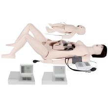
An Anatomical Model of a dog uterus typically includes the following features: 1. Two horns: The dog uterus is a bicornuate organ, meaning it has two horns that extend from the body of the uterus. These horns are where the puppies develop during pregnancy. 2. Body of the uterus: The body of the uterus is the central portion of the organ where the horns join together. This is where the fertilized eggs implant and develop into embryos. 3. Cervix: The cervix is the narrow passage that connects the uterus to the vagina. It helps to keep the uterus closed during pregnancy and opens during labor to allow the puppies to pass through.
4. Ovaries: The ovaries are the female reproductive organs that produce eggs (ova) and hormones. They are located near the ends of the uterine horns.
5. Fallopian tubes: The fallopian tubes are the ducts that connect the ovaries to the uterus. They transport the eggs from the ovaries to the uterus for fertilization. An anatomical model of a dog uterus can be used for educational purposes, such as teaching veterinary students about the reproductive system of female dogs. It can also be helpful for pet owners to understand the anatomy of their dog's reproductive system and the importance of spaying or neutering to prevent unwanted litters.
Parameter:Showing the vagina, uterus, uterine horns, ovaries