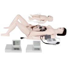
An Anatomical Model of a pig uterus would typically include the following features: 1. Uterine horns: The pig uterus has two long, curved structures called uterine horns that extend from the body of the uterus. These horns are where fertilized eggs implant and develop into embryos. 2. Body of the uterus: The body of the uterus is the central portion of the uterus where the uterine horns converge. This is where the embryos develop before being expelled during parturition. 3. Cervix: The cervix is the narrow, muscular opening at the end of the uterus that leads to the vagina. It helps to keep the uterus closed during pregnancy and opens during parturition to allow the passage of the fetus. 4. Ovaries: The ovaries are small, oval-shaped organs located near the uterine horns. They produce eggs (ova) and hormones such as estrogen and progesterone that regulate the reproductive cycle.
 5. Ligaments: The uterus is supported by several ligaments that help to hold it in place within the abdominal cavity. These ligaments also provide stability and allow for movement of the uterus during pregnancy.
 An anatomical model of a pig uterus can be used for educational purposes in veterinary and biology classes to help students understand the structure and function of the reproductive system in pigs. It can also be used for research purposes to study reproductive disorders and diseases in pigs.
Parameter:Showing the uterus, uterine horns, and fetus