The kidney, an essential organ responsible for filtering blood and removing waste products, relies on the proper functioning of its components such as the nephron and glomerulus. Understanding the structure and function of these components is crucial for maintaining kidney health and diagnosing kidney conditions.
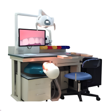
In medical training, various models and manikins are used to simulate real-life scenarios and practice different skills. For example, first aid skill training models, such as the " Full Body Trauma Manikin " and " Mattress Sutures Model ," provide realistic training for emergency situations. Additionally, models like the " Intravenous Injection Arm " and " Intramuscular Injection Model " help healthcare professionals practice precise injection techniques.
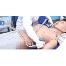
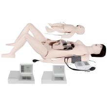
It is important to have specialized training models for specific medical fields. Obstetrics And Gynecology Skills can be developed with models like the "Advanced Maternity Examination Model" or the "Uterine Curettage Training Model." Similarly, Surgical Skills can be honed using models like the "Rectal Examination Training Model" or the "Intestine Surgical Procedure Model."
Nursing skills require hands-on practice, and models like the " Adult Nursing Intravenous Arm Model " and "Monitoring Female Catheterization Model" offer a realistic learning experience. Pediatric care can be simulated using models such as the "Interactive Infant Simulation Model" and "Newborn Physical Examination Model."
Different anatomical models assist in understanding the human body. These models include the " Anatomical Human Brain Model " for studying the Nervous System , the " Urinary System Model " for comprehending the kidney's structure, and the " Human Muscular System Model " for learning about muscles.

Overall, the utilization of training models and manikins enhances medical education by providing a safe and practical environment for healthcare professionals to develop their skills and knowledge in various medical fields.
Features:
Model consists of 3 models, demonstrates structure of the renal section plane (include cortical substance, medulla, proximal convoluted tubule, distal conveoluted, connecting canal, mammillary ducts, minor renal calices, greater renal calices, pelvis of ureter, ureter),nephron structure, golomerus (include renal capsule, glomus) and blood vessels.