This Anatomical Model provides a comprehensive and detailed depiction of the pancreas, spleen, and duodenum, offering a unique opportunity for a thorough examination of their intricate structures and vital functions. It serves as an invaluable tool for medical students and healthcare professionals, especially those undergoing Imaging Medicine Model Training.
One of the key features of this model is the openness of the pancreas, allowing for a clear view of the entire pancreas duct. This duct is responsible for the transportation of essential digestive enzymes and hormones throughout the body, ensuring proper digestion and metabolic processes. By showcasing the pancreas duct, this model enhances the understanding of its crucial role in maintaining overall bodily health, a key aspect in Diagnostic Skills training.
In addition to the pancreas, the duodenum, the initial segment of the small intestine, is partially dissected to expose its inner structure. This includes the presence of villi and crypts, which are responsible for the absorption of nutrients from the food we consume. The model provides a deeper insight into the process of nutrient absorption and its significance in sustaining our body's energy and overall well-being, a crucial part of Viscerology studies.
Furthermore, the inclusion of the spleen in this anatomical model is of great importance. The spleen is a vital organ of the immune system, playing a crucial role in filtering the blood and removing old or damaged red blood cells. By featuring the spleen, this model emphasizes the interconnection between the immune system and other organ systems, enhancing the understanding of the body's overall defense mechanisms. This is vital for those pursuing Trauma Care training.
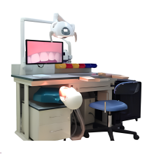
 Overall, this detailed representation of the pancreas, spleen, and duodenum is an excellent resource for gaining a deeper understanding of the complexities of the human body. Its life-sized display and intricate details offer an immersive learning experience, allowing for a comprehensive exploration of these organs' structure and function. Whether used for educational purposes or personal curiosity, this model, similar to an Ultrasound Guided Puncture Simulation Table, serves as a practical tool for learning and understanding.
Features:
Life size, pancreas, spleen and duodenum are displayed, the pancreas is open to show the entire pancreas duct, and duodenum is partially dissected to expose inner structure.


 Overall, this detailed representation of the pancreas, spleen, and duodenum is an excellent resource for gaining a deeper understanding of the complexities of the human body. Its life-sized display and intricate details offer an immersive learning experience, allowing for a comprehensive exploration of these organs' structure and function. Whether used for educational purposes or personal curiosity, this model, similar to an