A Thoracic Cavity Closed Drainage Model is a representation or simulation of the mechanics and functions of draining fluid or air from the thoracic cavity, which is the space within the chest that contains the lungs, heart, and other vital organs.
This model typically includes a detailed depiction of the thoracic cavity, including its anatomical structures, such as the ribs, diaphragm, and pleura (the thin membrane that lines the chest cavity and covers the lungs). It also incorporates a simulation of the fluid or air that needs to be drained from the cavity.
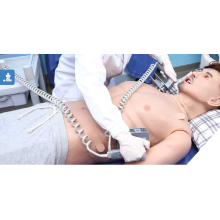
The model may simulate various scenarios, such as a pleural effusion (an accumulation of fluid in the chest cavity) or a pneumothorax (the presence of air in the chest cavity). It would represent the methods used for drainage, which often involve the insertion of a chest tube, catheter, or other devices to remove the excess fluid or air.
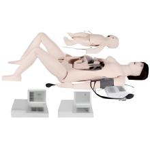
One essential aspect of the thoracic cavity closed drainage model is its ability to provide realistic training for medical professionals. For example, it can be combined with first aid skill training models, such as an Intravenous Injection Arm or a Mattress Sutures Model, to create a comprehensive training experience. Medical practitioners can practice their skills in inserting drainage devices, performing necessary procedures, and managing complications using high fidelity simulation models like the Full Body Trauma Manikin.
Additionally, the drainage model can be integrated into advanced medical training programs like ACLS (Advanced Cardiovascular Life Support) training. This allows healthcare providers to learn and practice life-saving techniques, such as CPR (cardiopulmonary resuscitation), using electronic CPR manikins or full-body Cpr Training manikins.
The thoracic cavity closed drainage model can also be used for specialized training in areas such as Tracheal Training, airway anatomy, gastric lavage, hemostasis, and more. By providing a realistic and interactive simulation, it enables healthcare professionals to enhance their Diagnostic Skills, develop proficiency in various procedures, and gain a better understanding of complex medical conditions.
It is important to note that the use of such models should be accompanied by proper supervision and guidance from experienced instructors. These models serve as valuable tools for education and training, but they cannot replace real-life clinical experience and expertise. However, through the integration of technology and anatomical accuracy, these models contribute significantly to the development of proficient and knowledgeable healthcare professionals.
Features:
1. Clear anatomical structure for convenient locating
2. Left side is operation area, right side is demonstration area, demonstrate structure of thoracic cavity
3. Allow repeating operation
4. Thoracic cavity closed drainage operation
5. Treatment after drainage
6. Pneumothorax puncture
7. Extracting pleural effusio