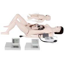
The Anatomical Model of a bovine uterus typically consists of a life-sized replica of the reproductive organ of a cow. It is often made of durable, high-quality materials such as plastic or silicone to accurately represent the structure and features of the uterus. The model typically includes the following parts: 1. Uterine horns: The two long, curved structures that extend from the body of the uterus and connect to the ovaries. These are where fertilization and early embryo development occur. 2. Body of the uterus: The central portion of the uterus where the fertilized egg implants and grows into a fetus during pregnancy. 
3. Cervix: The lower part of the uterus that connects to the vagina and serves as a passageway for sperm to enter the uterus and for the fetus to exit during birth.
4. Ovaries: Small, oval-shaped organs located near the ends of the uterine horns that produce eggs (ova) and hormones essential for reproduction. An anatomical model of a bovine uterus is often used in veterinary education, research, and reproductive health training to help students and professionals understand the anatomy and function of the reproductive system in cows. It can also be used for demonstrating artificial insemination techniques, embryo transfer procedures, and other reproductive technologies.
Parameter:Cross section of vagina, uterus, and uterine horn