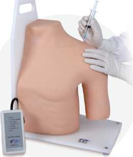
A shoulder joint cavity injection model is a three-dimensional representation of the shoulder joint and surrounding structures. It is used to demonstrate and practice the injection technique for administering medication or other substances into the shoulder joint cavity.
The model typically includes the humerus bone, scapula bone, and the surrounding ligaments, tendons, and muscles of the shoulder joint. It also includes a simulated joint cavity, which is the space between the humerus and scapula where the injection is performed.
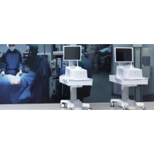
Shoulder Joint Cavity Injection Model
The purpose of the First Aid Skill Training Model is to provide a realistic and anatomically accurate representation of the shoulder joint, allowing healthcare professionals to practice and improve their injection technique. It can be particularly useful for medical students, residents, and healthcare professionals who need to administer injections into the shoulder joint cavity as part of their practice.
By using the Intravenous Injection Arm model, healthcare professionals can familiarize themselves with the anatomy of the shoulder joint, identify the correct injection site, practice proper needle insertion and angle, and develop their skills in a controlled and safe environment before performing the procedure on a patient.
Overall, the Mattress Sutures Model is a valuable tool for training and education in the field of healthcare, helping to improve the accuracy and safety of shoulder joint injections.
Features:
1. Precise anatomical structure, shoulder blade, clavicle, humerus, the deltoid, the biceps brachii and shoulder joint ligament
2. Practise shoulder joint puncture
3. 6 puncture positions:
1) Shoulder joint cavity (front)
2) Shoulder joint cavity (rear)
3) Subacromial bursa
4) Shoulder lock joint
5) Tendon sheath
6) Nerve block on the shoulder
4. Electronic monitoring with light indication